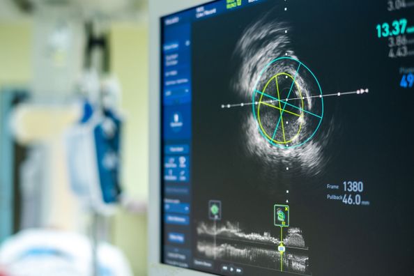
Throughout the decades healthcare has continued to advance in technology, customer service and treatment options to support and improve preventive care, disease management and recovery for patients. Cardiology in particular has seen groundbreaking innovations in advanced testing and minimally invasive procedures to help identify and treat cardiovascular disease early.
With heart disease being the leading cause of death for both men and women in the United States, it is essential that imaging, procedures and preventive care health practices continue to grow and evolve with recent innovations.
Advanced cardiac imaging is the foundation for discovering accurate and early diagnosis. Through precise imaging, cardiologists are able to identify your heart condition and create an effective treatment plan for your diagnosis. The following imaging techniques have become some of the most effective methods for identifying heart disease.
Nuclear Cardiac Imaging
Advanced nuclear imaging for cardiovascular disease produces accurate, high quality images of your heart and blood flow. During this test, a small dose of radioactive tracer is injected into your blood stream and travels through the area that is being examined. The tracer emits gamma rays, which are detected by a camera, that will take images to map blood flow and evaluate heart muscle function and your coronary arteries. The test will be performed in a medical lab, and depending on the type of image your physician is looking to obtain, he/she will order a computed tomography (CT) scan, positron emission tomography (PET) scan or magnetic resonance imaging (MRI) that can produce special views specific to your heart condition.
These tests allow our cardiologists to identify significant heart conditions, and/or obstructive blockages in your coronary circulation, that may require angioplasty (surgical blood vessel repair) or open-heart bypass surgery to restore normal blood flow in your heart. With tens of thousands of nuclear imaging studies already performed, our technologists and cardiologists are able to critically evaluate and offer their expert advice on a treatment plan for your specific heart condition.
CardioMEMs
CardioMEMS is a heart failure monitoring system that provides our cardiologists with pulmonary artery pressure monitoring and hemodynamic data to proactively manage your heart failure condition. Your physician will be able to use the CardioMEMs device information to make changes to your treatment before you feel symptoms. The wireless device is implanted using a catheter based procedure and does not require a battery or any replaceable parts.
Echocardiography
An echocardiography (echo) is a pain-free test that uses sound waves to create moving pictures that show the size and shape of your heart. With constant advancements in technology, the echo has evolved into many test forms that can be used to produce cardiac results specific to what your physician is examining.
The most common type of echo is called the transthoracic echocardiography. During this test, a device called a transducer is placed on your chest, sending ultrasound waves through your chest wall to create a moving picture of your heart. The transthoracic echo is used to examine problems with heart valves and blood flow. The results from this test can help your cardiologist identify the amount of blood being pumped out of the left ventricle, valve function and areas of poor blood flow in the heart, as well as assessment of previous heart injuries.
The echo can also be used as a part of cardiac stress testing. During a stress test, your cardiologist will have you exercise or take medicine to produce a hard and fast heartbeat. The echo transducer will start imaging your heart while it is working harder than normal. An echo stress test is ordered by your cardiologist when you are experiencing chronic chest pain that may be due to coronary artery disease. The stress test is able to determine the cause of chest pain as well as how much exercise you can safely tolerate during cardiac rehabilitation.
One of the most significant advancements of the echo is the development of three-dimensional (3D) imaging. Real-time 3D imaging allows for improvement in the accuracy and visualization of cardiac valves and congenital abnormalities that can provide cardiologists with more guidance on interventions for heart disease. This test is performed like the transthoracic echo, but uses a higher quality imaging technique. The 3D echo can help identify significant heart conditions and improper blood flow and can also be used to oversee heart function during surgery as well as monitor an un-born baby’s heartbeat.
As advanced imaging techniques continue to evolve and are available to patients with heart conditions, they help increase early diagnosis and preventive health treatments that can lower your risk of chronic heart disease or failure.
For a full list of our cardiology imaging techniques, please visit our cardiology page. For more information or to schedule an appointment with a cardiologist, schedule an appointment online or by calling your preferred location.
Health Topics:

